Figure 5
RBD targeted COVID vaccine and full length spike-protein vaccine (mutation and glycosylation role) relationship with procoagulant effect
Luisetto M*, Tarro G, Farhan Ahmad Khan, Khaled Edbey, Mashori GR, Yesvi AR and Latyschev OY
Published: 26 April, 2021 | Volume 5 - Issue 1 | Pages: 001-008

Figure 5:
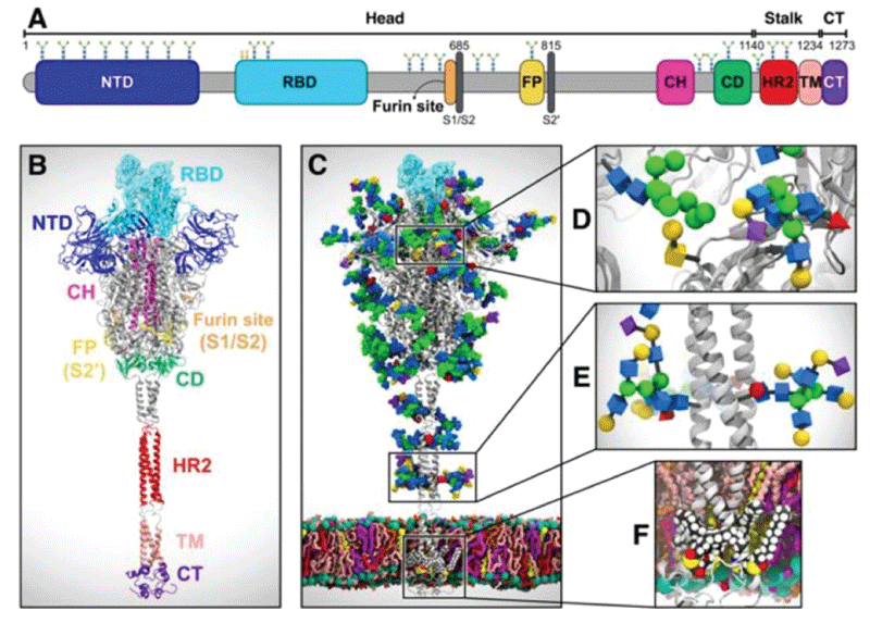
System overview. (A) A sequence of the full-length spike (S) protein contains the N-terminal domain (NTD, 16-291), receptor binding domain (RBD, 330-530), furin cleavage site (S1/S2), fusion peptide (FP, 788-806), central helix (CH, 987-1034), connecting domain (CD, 1080-1135), heptad repeat 2 (HR2, 1163-1210) domain, transmembrane domain (TD, 1214-1234), and cytoplasmic tail (CT, 1235-1273). Representative icons for N-glycans (blue and green) and O-glycan (yellow) are also depicted according to their position in the sequence. (B) Assembly of the head, stalk, and CT domains into a full-length model of the S protein. (C) Fully glycosylated and palmitoylated model of the Open system. (D-F) Magnified view of the N-/O-glycans rendered using the Symbol Nomenclature for Glycans (SNFG) (D, E) and S-palmitoylation of the cytoplasmic tail (F). from Casalino, et al.
Read Full Article HTML DOI: 10.29328/journal.jcavi.1001007 Cite this Article Read Full Article PDF
More Images
Similar Articles
-
RBD targeted COVID vaccine and full length spike-protein vaccine (mutation and glycosylation role) relationship with procoagulant effectLuisetto M*,Tarro G,Farhan Ahmad Khan,Khaled Edbey,Mashori GR,Yesvi AR,Latyschev OY. RBD targeted COVID vaccine and full length spike-protein vaccine (mutation and glycosylation role) relationship with procoagulant effect. . 2021 doi: 10.29328/journal.jcavi.1001007; 5: 001-008
Recently Viewed
-
The Lived Experiences of Addiction Counselors and Applications for Building ResiliencyBrian N Paulson*, Amy L Hayes, Stacey C Lilley, Brad A Imhoff, Charlotte A Crosland*. The Lived Experiences of Addiction Counselors and Applications for Building Resiliency. J Addict Ther Res. 2024: doi: 10.29328/journal.jatr.1001031; 8: 024-034
-
Changes in Serum Markers of Atherogenesis and Hematological Profile after the consumption of Quail eggsEkpenyong CE*,Patrick ES,Akpan KS. Changes in Serum Markers of Atherogenesis and Hematological Profile after the consumption of Quail eggs. Arch Food Nutr Sci. 2017: doi: 10.29328/journal.afns.1001001; 1: 001-011
-
Generating eco-friendly electricity from rain waterKhalid Abdel Naser Abdel Rahim*. Generating eco-friendly electricity from rain water. Ann Civil Environ Eng. 2022: doi: 10.29328/journal.acee.1001032; 6: 001-002
-
Percutaneous Closure of Post-myocardial Infarction Ventricular Septal Rupture-experience From a Resource-limited Setup From Eastern Part of IndiaSiddhartha Saha*. Percutaneous Closure of Post-myocardial Infarction Ventricular Septal Rupture-experience From a Resource-limited Setup From Eastern Part of India. J Cardiol Cardiovasc Med. 2024: doi: 10.29328/journal.jccm.1001184; 9: 087-092
-
Value of Speckle Tracking Echocardiography in Prediction of Left Ventricular Reverse Remodeling in Patients with Chronic total Occlusion Undergoing Percutaneous Coronary InterventionsGehan Magdy*, Sahar Hamdy Azab, Yasmin Ali Esmail, Mohamed Khalid Elfaky. Value of Speckle Tracking Echocardiography in Prediction of Left Ventricular Reverse Remodeling in Patients with Chronic total Occlusion Undergoing Percutaneous Coronary Interventions. J Cardiol Cardiovasc Med. 2023: doi: 10.29328/journal.jccm.1001170; 8: 164-170
Most Viewed
-
Evaluation of Biostimulants Based on Recovered Protein Hydrolysates from Animal By-products as Plant Growth EnhancersH Pérez-Aguilar*, M Lacruz-Asaro, F Arán-Ais. Evaluation of Biostimulants Based on Recovered Protein Hydrolysates from Animal By-products as Plant Growth Enhancers. J Plant Sci Phytopathol. 2023 doi: 10.29328/journal.jpsp.1001104; 7: 042-047
-
Sinonasal Myxoma Extending into the Orbit in a 4-Year Old: A Case PresentationJulian A Purrinos*, Ramzi Younis. Sinonasal Myxoma Extending into the Orbit in a 4-Year Old: A Case Presentation. Arch Case Rep. 2024 doi: 10.29328/journal.acr.1001099; 8: 075-077
-
Feasibility study of magnetic sensing for detecting single-neuron action potentialsDenis Tonini,Kai Wu,Renata Saha,Jian-Ping Wang*. Feasibility study of magnetic sensing for detecting single-neuron action potentials. Ann Biomed Sci Eng. 2022 doi: 10.29328/journal.abse.1001018; 6: 019-029
-
Pediatric Dysgerminoma: Unveiling a Rare Ovarian TumorFaten Limaiem*, Khalil Saffar, Ahmed Halouani. Pediatric Dysgerminoma: Unveiling a Rare Ovarian Tumor. Arch Case Rep. 2024 doi: 10.29328/journal.acr.1001087; 8: 010-013
-
Physical activity can change the physiological and psychological circumstances during COVID-19 pandemic: A narrative reviewKhashayar Maroufi*. Physical activity can change the physiological and psychological circumstances during COVID-19 pandemic: A narrative review. J Sports Med Ther. 2021 doi: 10.29328/journal.jsmt.1001051; 6: 001-007

HSPI: We're glad you're here. Please click "create a new Query" if you are a new visitor to our website and need further information from us.
If you are already a member of our network and need to keep track of any developments regarding a question you have already submitted, click "take me to my Query."


